What is Uveitis?
Uveitis is the inflammation of the uvea (vascular middle layer of the eye). Uvea is divided into 3 layers in particular, from front to back: i) Iris ii) Ciliary body iii) Choroid. It lies between the inner retina and the outer fibrous layer composed of the sclera and cornea. Uveitis is an eye-related disease and requires a thorough examination by an ophthalmologist or optometrist and urgent treatment to control the inflammation. As its warning signs often come on suddenly and get worse quickly.
Causes:
Certain autoimmune disorders can lead to Uveitis. Some of them are rheumatoid arthritis, ankylosing spondylitis, psoriasis, arthritis.
Eye injury or surgery, An infection, such as cat-scratch disease, herpes zoster, syphilis, toxoplasmosis, or tuberculosis
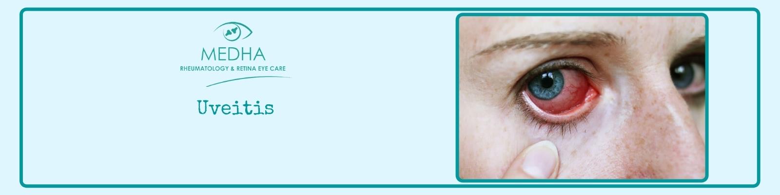
Signs and Symptoms:
Uveitis gives different indications to different layers of the Uvea.
Burning of the eye, Eye redness, Blurred vision, Photophobia, Irregular pupil, dilated ciliary vessels, and Busacca nodules are the signs of anterior uveitis(iritis).
Floaters and Blurred vision are signs in Intermediate uveitis and these signs also present in Posterior uveitis.
Panuveitis arises when all layers of the uvea are inflamed, from the front to the back of your eye.
Diagnosis:
Laboratory testing is usually used to diagnose particular underlying diseases, including rheumatologic tests (e.g. antinuclear antibody, rheumatoid factor) and serology for infectious diseases.
Dilated fundus examination- is a diagnostic method that requires the use of mydriatic eye drops (such as tropicamide) to dilate or enlarge the pupil to obtain a better view of the fundus of the eye. It is used to rule out posterior uveitis, which cites white spots across the retina along with retinitis and vasculitis.
Fluorescein angiography or indocyanine green angiography. These tests require the placement of an intravenous (IV) catheter in a vein in your arm to administer a dye. This dye will reach the blood vessels in the eyes and allow photographs of blood vessel inflammation inside the eyes.
A tonometry exam scales the pressure inside your eye (intraocular pressure). Numbing eye drops may be used for this test.
Treatment:
The goal of treatment is to reduce the inflammation in your eye, as well as in other parts of the body if present.
Uveitis is typically treated with glucocorticoid steroids, either as topical eye drops (prednisolone acetate) or as oral therapy.
Antimetabolite medications, such as methotrexate are frequently used for recalcitrant or more severe cases of uveitis.
Vitrectomy- Surgery to remove some of the vitreous in your eye is rarely used to diagnose or manage the condition.
A medication-releasing implant- A device that's implanted in the eye may be an option, for people with difficult-to-treat posterior uveitis. This device slowly releases corticosteroid into the eye for two to three years.
Prevention:
Seeking proper treatment for an autoimmune disease or infection can help to prevent uveitis.
Early detection and treatment are important to reduce the risk of vision loss, which can be permanent.